Friday, 21 March 2014

Brain stroke treatment in india, Cost Brain Stroke Treatment in India, Brain stroke surgery in India

A stroke occurs when blood supply to the brain is interrupted or there is bleeding in the brain. Within minutes, brain cells begin to die. It is urgent to get emergency care at the first sign of a stroke. Early treatment saves many lives and reduces the effects of stroke.
Stroke Causes Of the 2 main types of stroke, the ischemic stroke occurs 80-85% of the time. With an ischemic stroke, a blood vessel in the brain becomes clogged. With a hemorrhagic stroke, a blood vessel in the brain actually bursts or leaks.


Hemorrhagic stroke


Hemorrhagic strokes tend to be more serious. The distinction between type of stroke can be critical in determining the treatment used.

   * Ischemic strokes occur when a blood vessel gets so narrow or clogged that not enough blood can get through to keep the brain cells alive.
   * Hemorrhagic strokes occur when the wall of a blood vessel becomes weak and blood leaks out into the brain.


What are the types surgery for brain stroke treatment in India?

After initial emergency management of stroke there might be the cases that may require surgery for treatment of stroke. Option of Surgery for treatment of stroke in India is available in following cases:

Surgery for Brain Stroke Due to Blockages in Blood Vessel in Brain: Most brain strokes occur when a blood vessel in the brain is blocked and blood flow stops. This type of stroke is called an ischemic stroke. The blockage may be caused by a blood clot, and severe brain swelling may result. Life-saving surgery in this type of stroke may be necessary as a better treatment to remove the clot and the brain tissue that has died from lack of oxygen.

Surgery for Brain Stroke Due to Blockage in main artery in the Neck:The main arteries in the neck help supply the brain with blood. They are called the carotid arteries. When patients have a serious blockage in these arteries, surgery may be done to prevent a stroke or a ministroke, which is also called a TIA. The operation is called a carotid endarterectomy.


Surgery for Brain Stroke Due toAltered Blood Flow: Blood flow to the brain may decrease temporarily in some patients. This is called a ministroke
or a TIA, which stands for Transient Ischemic Attack. While the brain is not getting enough blood, it cannot work properly. Patients who have TIAs get symptoms for a short time that make it difficult for them to function. Bypass surgery may be advised for some patients who continue to have TIAs.


Surgery for Brain Stroke Due to Internal Bleeding: Bleeding in the brain causes some strokes. These strokes are called hemorrhagic stroke. The bleeding may occur when a weakened blood vessel leaks or bursts. This is called an aneurysm. When an aneurysm occurs, the weakened artery may become like a balloon filled with blood. Patients usually describe an aneurysm as the worst headache of their life.


There are several types of surgery to repair an aneurysm. A clip may be placed across the neck of the aneurysm (like a clip at the end of a balloon) to stop the bleeding. A newer approach is to thread a long, thin tube through the artery that leads to the aneurysm. Then a tiny coil is fed through the tube into the aneurysm “balloon” to fill the space and seal off the bleeding. 

Treating a stroke depends on where the stroke occurred in your brain and whether it's ischemic or hemorrhagic. The doctor may use a magnetic resonance imaging (MRI) scan, computed tomography (CT) imaging or angiography (injecting dye through a catheter inserted into the suspected blocked blood vessel and taking x-rays of the vessels) to determine the stroke type and location.

Time is critical. Until a few years ago, strokes were regarded as untreatable. Brain cells were thought to die within minutes after a stroke began, so stroke treatment was believed useless. The only onsite medical treatment is stabilization and "wait and see." Now researchers have discovered that some brain cells die immediately after a stroke, but others can survive for as long as several days. It is now clear that treatment following a stroke, especially if begun within three hours of onset, can help preserve brain tissue.


For more information visit:          http://www.medworldindia.com        
                    
https://www.facebook.com/medworld.india

Please scan and email your medical reports  to us at care@medworldindia.com and we shall get you a Free Medical Opinion from India’s Best Doctors.

Call Us : +91-9811058159
Mail Us : care@medworldindia.com


Tuesday, 18 March 2014

The Advantage of Laminoplasty spine Surgery – Stenosis , Herniated Disc , Spondylosis , Tumor Treatment in India


Laminoplasty or laminaplasty is a term that refers to a surgical procedure performed to relieve spinal cord and/or nerve root compression.  To help you understand, let’s separate laminaplasty into two; lamina and plasty.
  • Lamina - Located at the back of the spine, the lamina are thin bony plates between each facet joint in the cervical (neck), thoracic (mid back), and lumbar (low back) spine.  The laminar plates are part of the bony wall that covers and protects the spinal canal.  Within the spinal canal are the spinal cord and nerve rootlets.
  • Plasty means to surgically repair.
Your spinous processes are bones that project off the back of your spine.  You can feel the end of most of your spinous processes by moving your hand up or down your spine.

Purpose

During laminaplasty, the lamina is cut in such a way that it opens like a door, and takes pressure (decompresses) off the spinal cord and nerve roots.  Your neurosurgeon uses small pieces of bone or metal plate as a wedge to hold the lamina in position during healing.

Causes of spinal cord and/or nerve root compression includes:
  • Cervical spinal stenosis
  • Cervical herniated disc
  • Cervical radiculopathy
  • Degenerative disc disease
  • Lumbar herniated disc
  • Lumbar spinal stenosis
  • Spondylosis
  • Spondylolisthesis
  • Tumor, infection (uncommon)\

Laminoplasty aims at decompressing the spinal cord and the spinal nerves, by hinging open the vertebrae posteriorly. The lamina (flat arch on the backside of the vertebral body) is cut open on one side and grooved on the other side to keep it hinged to the main body of the vertebra. This creates more room for the spinal cord and nerves. Any compressing structure like a herniated or fragmented disc, or bony spurs, thickened ligament etc are also removed in this procedure.

The advantage of this procedure is that the stability of the spine is maintained as the amount of bone and muscle tissue that is removed is very less, and any fusion surgery of the spine is avoided.

How is the surgery performed?

This surgery is performed by sedating the patient under general anesthesia and making him lie on his stomach. The head is kept slightly bent with the help of Mayfield clamp to straighten the skin folds on the neck. A midline incision is made on the back of the neck corresponding to the affected spinal segment. The skin is cut open and the muscles are separated to view the involved vertebrae.

The lamina are cut through their thickness longitudinally on one side and grooved on the other side to keep it hinged to the vertebral body. The posterior part of the vertebra is swung open like a door. Small wedges made of bone are placed in the opened space of the door. The door of the vertebrae swings shut, and the wedges stop it from closing all the way. The spinal cord and the nerve roots rest comfortably behind the door. Since this increases the space in the spinal canal, it decompresses the spinal cord with immediate relief of symptoms. Any presence of herniated disc, thickened ligament or facet joint and osteophytes are identified and removed.


The person is usually up and about on the same day after surgery and is discharged in one or two days later. The Physical therapist advises certain exercises of the neck to maintain the flexibility and strength of the neck muscles before the patient is discharged. Certain ergonomics advice regarding neck movements is also given.

The patient is able to return to his daily activities wearing a soft cervical collar within a few days. The collar is discarded after some time.

Our network hospitals have the most advanced spine surgery facilities. The spine surgeons here use minimally invasive and computer guided techniques in spine surgery. In a Minimally Invasive spine surgery the surgeon makes a few small incisions unlike the open surgery where a single large incision is made. Minimally Invasive spine surgery has several important benefits for the patients. If you have been advised spine surgery your first choice should be the latest minimally invasive technique developed by our world best hospitals in India. Most advanced Technology like Intra Operative MRI, Brain Suite and Computer Assisted Navigation System are deployed by highly trained surgeons for accurate and safe Spine Surgeries through a small incision.

Why should you choose to get Indian hospitals offer the best spinal surgery treatment in India at affordable prices. MedWorld india associated best spine surgery hospitals in India have the latest technology and infrastructure to offer the most advanced spine surgery at low cost.

Salient features of these hospitals are:
  • Comprehensive management of spine disorder from birth defects to degeneration of tumor and trauma.
  • Dedicated team of International trained and vastly experienced Spine Surgeons, Rheumatologists, Neurologist, Physicians and Physiotherapist.
  • Expert evaluation of spinal problems by dedicated team of experienced spine Surgeons, Rheumatologists, Neurologist, Physicians and Physiotherapist.
  • Latest Generation Diagnostic and Imaging facilities including dynamic digital X rays, Spiral CT scanning , MRI and Electrophysiology unit all under one roof.
  • Physiotherapy and Rehabilitation by experts after the surgery help you regain functional abilities quickly helping in vastly improved overall results.

 For more information visit:          http://www.medworldindia.com         
                    https://www.facebook.com/medworld.india

Please scan and email your medical reports  to us at care@medworldindia.com and we shall get you a Free Medical Opinion from India’s Best Doctors.

Call Us : +91-9811058159
Mail Us : care@medworldindia.com

Monday, 10 March 2014

Cervical Spinal Stenosis Treatment in India - About Back & Neck Pain

Most cervical problems are due to degenerative changes that occur in the discs and joints of the neck. Degenerative changes that affect the structures of the spine can cause the spinal canal to become too narrow, a condition called spinal stenosis. This may lead to pressure on the spinal cord. Bone spurs that stick into the spinal canal take up space, making the spinal canal smaller. They can press against the spinal cord or nerve roots.

Pressure on the spinal cord from spinal stenosis can cause symptoms of myelopathy. Myelopathy may impair normal walking, hand and finger use, and bowel and bladder function. Doctors take these symptoms very seriously because severe myelopathy that is not treated may lead to permanent nerve or spinal cord damage. Pressure on nearby nerve roots can cause radiculopathy and may produce pain, weakness, or sensory changes in the area supplied by nerves that go from the cervical spine to the shoulder, arm, or hand.


Epidural Steroid Injection (Nerve Block)
If other treatments do not relieve your neck pain, you may be given an epiduralsteroid injection (ESI), also called a nerve block. An ESI places a small amount of cortisone into the bony spinal canal. Cortisone is a strong anti-inflammatory medicine that may control the inflammation surrounding the nerves and may ease the pain caused by irritated nerve roots. This injection is often used when other conservative measures do not work, or in an effort to postpone surgery. This treatment is not always successful but may provide short-term help. Learn more about spinal injections.

Physical Therapy
If your condition is causing only mild symptoms and does not appear to be getting worse, your doctor may have you work with a physical therapist. A well-rounded rehabilitation program assists in calming pain and inflammation, improving your mobility and strength, and helping you do your daily activities with greater ease and ability.

In mild cases, nonsurgical treatments offer ways to control symptoms and enable you to improve without surgery. You may be issued a neck brace to hold your neck steady, reducing extra strain. You will need to limit your activities, especially heavy and repeated movements of the arms and neck. Therapists use electrical stimulation to ease muscle spasm and pain. Treatments may also include cervical traction to gently stretch the joints and muscles in the neck. Therapy sessions may be scheduled two to three times each week for up to six weeks.

The goals of physical therapy are to help you
·         learn correct posture and body movements to minimize the           effects of stenosis
·         maintain appropriate activity levels
·         identify symptoms of stenosis that require medical attention
·         learn ways to manage your condition

Surgical Treatment

Cervical myelopathy can be a serious problem. The pressure on the spinal cord usually will not go away without surgery, and the symptoms may continue to get worse. If you do not improve with non-operative care, your doctor may suggest surgery.
There are several surgical procedures used to treat cervical spinal stenosis that is causing cervical myelopathy; all have the same goal-to relieve the pressure on the spinal cord by making the spinal canal larger.

Your surgeon may suggest an operation called a laminectomy, in which the backside of the vertebrae is opened to allow more room for the spinal cord. Discectomy, the removal of one or more discs, may be suggested if stenosis is coming from problems of disc herniation. This surgery may be done from the front or back of the spine. Or your surgeon may suggest an operation that is done from the front of the neck-a corpectomy and strut graft. This operation involves removing the discs and vertebral bodies in the area where problems are occurring. Bone spurs that are pushing into the spinal cord are also removed. The vertebrae are then replaced with a solid piece of bone graft (called a strut graft). The strut graft heals over time to create a solid fusion of the spine where the vertebral bodies have been removed.

World's Most Advanced Medical Treatment in India - Get free Expert Medical Opinion and Treatment Estimate Cost

MedWorld India offer free, no obligation assistance to international patients to find world class medical treatment in India. A large number of people from all over the world are now traveling to India for top class medical treatment like Heart Surgery, Cancer Care, Spinal fusion surgery in India, sleeve gastrectomy surgery in India, and other major surgeries. India offers an unmatched cost and quality advantages because it has world class hospitals and globally trained and experienced surgeons across every specialty. Whether you are considering cervical disk replacement surgery, brain tumor surgery in India, heart valve replacement surgery, or prostate cancer treatment in India, we offer support and services to facilitate the care you require. 
We can help you find the best heart hospital in India, IVF hospital in India, or best cancer hospital in India.
The First step is to email your Medical Reports to us for an opinion from leading doctors in India. We will send you an expert medical opinion and estimate for the cost of your treatment from at least three leading hospitals in India. Once you decide, we help you schedule appointments, apply for a medical Visa and make the arrangements for your stay.

MedWorld India has a team of dedicated doctors who personally attend to all your queries. We are the only facilitators that appoint a personal doctor to the patient from the time of enquiry till the time the patient fully recovers. Your personal doctor will stay in constant contact with you, your local doctor and your medical team in India throughout your stay, and will relay information back to your loved ones. From initial registration of interest, to returning the patient back to his or her home country after the procedure and recuperation, MedWorld India will ensure the whole process is as smooth and stress-free as possible for its patients. We believe that timely, affordable and quality medical treatment is every human beings right. We are dedicated to this cause and strive to deliver the benefit and pleasure of medical care to people across the globe.
Our mission at MedWorld India is simple. We want to help you to safely and successfully receive your medical procedure(s) at a world-class healthcare facility for a fraction of the cost elsewhere.

Advantages of MedWorld India

 Top Quality Healthcare services at Low cost
 Save up to 60-80% on medical costs
 Highly Qualified Physicians/Surgeons and Hospital support staff
 Get free Opinion - No charge for consultation
 Guide you to select the Best Hospital and Doctor for your procedure.
 Make all your arrangements for travel

Website: www.medworldindia.com
Email: care@medworldindia.com
Telephone: +91-9811058159
Facebook: https://www.facebook.com/medworld.india
Twitter : https://twitter.com/medworld_india


Friday, 7 March 2014

Low Cost Minimally Invasive Spine Surgery in India

MINIMALLYINVASIVE SPINE SURGERY  - Preserves  musculoskeletal structures as the surgery involves small incisions, shortened hospital  stay and faster return to normal activities Procedures include endoscopic repair of herniated disc, spinal decompressions and fusions, deformity correction (listhesis and scoliosis), minimally Invasive corpectomies  and tumor removal, percutaneous bone augmentation in osteoporosis and fractures (Vertebroplasty) and percutaneous spinal stabilization. Advanced telescopes (endoscopes) & Instrumentation

There is a rapid increase in technology that allows the MIS physician to treat patients with minimally invasive spine surgery procedures. Some of these techniques are now well established, while others remain new and continue to evolve. The best MIS (Minimally Invasive Spine Surgery) technique will depend on the specific character of the problem. While the list of MIS (Minimally Invasive Spine Surgery) techniques continues to expand, the most commonly employed today include:

WORLD CLASS TECHNOLOGY - The latest technology has a major role to play in diagnosing and assisting the advanced surgical procedures.

INTRA OPERATIVE MRI SURGERY SUITE- is an advanced operating theatre with capability of intra-operative MR imaging and MR guided surgery. Surgeon can perform a high resolution MRI during the surgery to assess the degree of tumor removal as well as to avoid normal tissues.

BIPLANE NEUROENDOVASCULAR ANGIOGRAPHY - Latest flat panel Biplane DSA (digital subtraction laboratory) provides ultrahigh resolution (2048 matrix) of cranial and spinal blood vessels. This unit is one of the most advanced units installed in this part of the world. The features include.

ADVANCED DYNA CT  and S - 7 NAVIGATION SYSTEM : the angiographic suite has a built in CT scan like facility for early detection of any complication.  Latest generation of Navigational equipment with integrated minimal access capabilities allows Real-time Navigational and imaging ability for better visualization of normal anatomy and the disease process allows preservation of normal anatomical structures and completeness of removal of the disease.


  • Neurologists – who diagnose and treat conditions of the nervous system focusing on specific area such as stroke, epilepsy,headache, movement disorders and other diseases.
  • Neurosurgeons - - having specific areas of interest such as brain tumor, spine surgery, endoscopic surgery, endovascular surgery, epilepsy surgery, Deep Brain Stimulation (DBS)
  • Intervention Neuroradiologists - use a nonsurgical, catheter based approach – similar to cardiac catheterization for heart disease – to treat vascular (blood vessel) diseases that affect the nervous system
 Our network hospitals have the most advanced spine surgery facilities. The spine surgeons here use minimally invasive and computer guided techniques in spine surgery. In a Minimally Invasive spine surgery the surgeon makes a few small incisions unlike the open surgery where a single large incision is made. Minimally Invasive spine surgery has several important benefits for the patients. If you have been advised spine surgery your first choice should be the latest minimally invasive technique developed by our world best hospitals in India. Most advanced Technology like Intra Operative MRI, Brain Suite and Computer Assisted Navigation System are deployed by highly trained surgeons for accurate and safe Spine Surgeries through a small incision.


Why should you choose to get Indian hospitals offer the best spinal surgery treatment in India at affordable prices. MedWorld india associated best spine surgery hospitals in India have the latest technology and infrastructure to offer the most advanced spine surgery at low cost.


  • Comprehensive management of spine disorder from birth defects to degeneration of tumor and trauma.
  • Dedicated team of International trained and vastly experienced Spine Surgeons, Rheumatologists, Neurologist, Physicians and Physiotherapist.
  • Expert evaluation of spinal problems by dedicated team of experienced spine Surgeons, Rheumatologists, Neurologist, Physicians and Physiotherapist.
  • Latest Generation Diagnostic and Imaging facilities including dynamic digital X rays, Spiral CT scanning , MRI and Electrophysiology unit all under one roof.
  • Physiotherapy and Rehabilitation by experts after the surgery help you regain functional abilities quickly helping in vastly improved overall results.


WORLD’sLOWEST COST  SPINE SURGERY PACKAGES -  All the above spine services come at a very attractive cost which is a fraction of the cost of similar services anywhere else in America and Europe.  Most  surgery packages can be availed of in less than 10000 US Dollars and that includes stay in a private  room where a companion can stay with the patient, surgeon fee, medicines and consumables, nursing care, patient’s food and airport pick up etc. etc.

Wednesday, 26 February 2014

Most important success factor in fusion surgery - Spinal Fusion Surgery in India


Lumbar spinal fusion is a type of back surgery in which a bone graft is inserted in the spine so that the bones in a painful segment of the spine fuse together. The fusion aims to stop the motion at a vertebral segment, which should decrease the pain caused by the joint. After the surgery it will take several months (usually 3 to 6, but sometimes up to 18 months) before the fusion is set-up. This surgery has been improved over the last 10 to 15 years, allowing for better success rates, and shorter hospital stays and recovery time.

Indications and contraindications for spinal fusion
The vast majority of people with low back pain will not need fusion surgery and will be able to manage the pain primarily with physical therapy and conditioning. A fusion surgery may, however, be recommended for patients with:
  • Low back pain caused by degenerative disc disease that limits the patient's ability to function (after non-surgical treatments, such as physical therapy and medication, have failed)
  • Isthmic, degenerative or postlaminectomy spondylolisthesis
  • A weak or unstable spine (caused by infections or tumors), fractures, or deformity (such as scoliosis)

Before beginning the main part of a spinal fusion procedure, your surgeon will need to gather material for a bone graft, which is used later on to join the targeted spinal bones together. Depending on individual circumstances and your surgeon’s preferences, this graft material can come from bone harvested from one of your own hips or ribs, from bone harvested from a special donor cadaver, or from artificial materials such as plastics or ceramics.

Once the graft material is ready, your surgeon will make an incision and create an opening that exposes the site of the fusion. Potential locations of this incision include your abdomen, your back and the side of your neck or torso. After exposing the site of procedure and removing the spinal disc that sits between your spinal bones, your surgeon can place the bone graft in one of several ways. First, he can place the graft material directly into the empty space left by the removed spinal disc. Alternatively, he can place the graft material inside a device called a spacer or cage, then insert this device into the gap between your spinal bones. Your surgeon can also lay the graft material over the rear surfaces of the targeted spinal bones.
All of these techniques will lead to fusion of your bones and incorporation of the graft material by triggering your body’s natural healing process. Frequently, bone grafts are held in position by metal rods or plates and screws during this healing period.

Spinal Fusion Uses

Problems in your spinal column that can lead to a need for spinal fusion include a form of bone slippage called spondylolisthesis, debilitating forms of abnormal spinal curvature or deformity, and mechanical spinal instability caused by problems such as accidents or injuries. Surgeries that can produce enough spinal instability to require fusion as a follow-up procedure include complete or partial removal of a damaged spinal disc (discectomy); removal of all or part of the section of spinal bone that forms the back of the spinal canal (laminectomy); and widening of the gaps in your spinal column that allow nerves to pass through from your spinal cord (foraminotomy).

Spinal Fusion Benefits

The rigidity and stability provided by spinal fusion can prevent dangerous degrees of spinal motion, ease pain and other symptoms associated with unwanted or excessive spinal motion, and help prevent damage in the soft tissues situated near your spine. In the vast majority of cases, these benefits are either permanent or remain for extremely extended periods of time.


Fusion surgery success rates vary between 70% and 95%, and there are several factors that will impact the success rate of the surgery, including:
  • Spine fusion for conditions that arise from gross instability (e.g. isthmic or degenerative spondylolisthesis) tends to be more successful than surgery done for pain alone (e.g. degenerative disc disease).
  • Individuals with only one badly degenerated disc (especially L5-S1) but an otherwise a normal spine tend to fare better than those undergoing multilevel fusions. Fusion surgery is generally considered for one or possibly two levels, and multilevel fusions should be avoided except in cases of severe deformity.
  • Individuals who have significant disc degeneration usually find more pain relief from a fusion than those with only minor degeneration on the MRI scan (e.g. still have a tall disc).

The most important success factor in fusion surgery is confirming that a patient's back pain is truly caused by degenerative disc disease, rather than some other condition. This is done by a combination of a careful review of the patient's history, a physical exam, and diagnostic tests (such as x-ray and MRI), and/or possibly a discogram.

For more information visit:          http://www.medworldindia.com        
                    
https://www.facebook.com/medworld.india

Please scan and email your medical reports  to us at care@medworldindia.com and we shall get you a Free Medical Opinion from India’s Best Doctors.

Call Us : +91-9811058159
Mail Us : care@medworldindia.com

Tuesday, 18 February 2014

Laminotomy surgery in india at Affordable Cost - Bone Spurs, Bulging Disc, Herniated Disc, Pinched Nerve, Spinal Stenosis

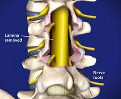
If spinal stenosis is the main cause of your neck pain, then the spinal canal must be made larger and any bone spurs pressing on the nerves must be removed. One way that this is done is with a complete laminectomy (lam-in-eck-toe-mee). Laminectomy means "remove the lamina". The lamina is the back side of the spinal canal and forms the roof over the spinal cord. Removing the lamina gives more room for the nerves and lets the surgeon remove any bone spurs from around the nerves. A laminectomy reduces the pressure on the spinal cord and relieves the irritation and inflammation of the spinal nerves.
In the cervical spine, removing the lamina completely may cause problems with the stability of the facet joints between each vertebra. If the joints are damaged during the laminectomy, the spine may begin to tilt forward, causing problems later. One way that spine surgeons try to prevent this problem is to not actually remove the lamina. Instead, they simply cut one side of the lamina and fold it back slightly. The other side of the lamina opens like a hinge. This makes the spinal canal larger giving the spinal cord more room. The cut area of the lamina eventually heals to keep the spine from tilting forward.

The traditional way of treating a herniated disc is to perform a laminotomy and discectomy. The term laminotomy means "make an opening in the lamina," and the term discectomy means "remove the disc."

This procedure is performed through an incision down the center of the back over the area of the herniated disc. The muscles are moved to the side so that the surgeon can see the back of the vertebrae. X-rays may be required during surgery to make sure the correct vertebra is located. The doctor cuts a small opening through the lamina bone on the back of the spinal column. This procedure, called "laminotomy," is used to give the doctor room to see and work inside the spinal canal. View animation of laminotomy.
The nerve roots are moved out of the way. Upon locating the problem disc, the surgeon removes it, easing pressure and irritation on the nerves of the spine. Small instruments that fit inside the disc are used to remove as much of the nucleus as possible. This prevents the remaining disc material from herniating in the future. View animation of discectomy. The muscles of the back are returned to their normal position around the spine. The skin incision is repaired with sutures or metal staples.

Microdiscectomy
Improvements have been made in the tools available to the spinal surgeon for performing a laminotomy and discectomy. Microdiscectomy is essentially the same as traditional discectomy, but this newer approach has several advantages. A much smaller incision is need when performing a microdiscectomy. There is less damage to nearby parts of the spine. Patients tend to recover faster.
A small incision is made in the back just above the area where the disc is herniated. Muscles are moved aside to see the vertebrae. The surgeon positions a microscope in the small incision. The remainder of the surgery is performed like the traditional method.

Endoscopic Discectomy
Many surgical procedures have been revolutionized by the use of special TV cameras. The procedure is still the same, but even smaller incisions (1/4 inch) are made to insert a special magnified TV camera into the spinal canal so that the surgeon can actually see the disc material. Through these tiny incisions, the camera and several other surgical instruments are inserted. Rather than looking through a microscope, the doctor watches the TV screen while working with specially designed instruments to remove the disc material.
Torn cartilages of the knee are now routinely removed with an arthroscope, and gallbladders are routinely removed with a laproscope. These small scopes give doctors a method of doing surgery with smaller incisions and with less risk to the nearby tissues. The same approach is evolving with spine surgery. Endoscopy of the spine is still in experimental stages and is not yet widely used. It might eventually give doctors a way to remove a disc with even less risk of injury than microdiscectomy.

A laminotomy of the spine is used to treat the following conditions:
This surgical procedure is carried out in two steps beginning with the laminotomy. Once this is accomplished, the second procedure, the micro discectomy, is performed. A high powered stereoscopic microscope is used to provide illumination and magnification to allow the nerve and surrounding structures to be visualized clearly through an incision less than one inch long. The nerve root is carefully protected with a specialized retractor, and protruding disc fragments, along with any remaining loose or degenerated disc material, are then removed with a small grasping device.


For more information visit:          http://www.medworldindia.com        
                    
https://www.facebook.com/medworld.india

Please scan and email your medical reports  to us at care@medworldindia.com and we shall get you a Free Medical Opinion from India’s Best Doctors.

Call Us : +91-9811058159
Mail Us : care@medworldindia.com


MedWorld India offer free, no obligation assistance to international patients to find world class medical treatment in India. A large number of people from all over the world are now traveling to India for top class medical treatment like Heart Surgery, Cancer Care, Spinal fusion surgery in India, sleeve gastrectomy surgery in India, and other major surgeries. India offers an unmatched cost and quality advantages because it has world class hospitals and globally trained and experienced surgeons across every specialty. Whether you are considering cervical disk replacement surgery, brain tumor surgery in India, heart valve replacement surgery, or prostate cancer treatment in India, we offer support and services to facilitate the care you require. We can help you find the best heart hospital in India, IVF hospital in India, or best cancer hospital in India.

The First step is to email your Medical Reports to us for an opinion from leading doctors in India. We will send you an expert medical opinion and estimate for the cost of your treatment from at least three leading hospitals in India. Once you decide, we help you schedule appointments, apply for a medical Visa and make the arrangements for your stay.

MedWorld India has a team of dedicated doctors who personally attend to all your queries. We are the only facilitators that appoint a personal doctor to the patient from the time of enquiry till the time the patient fully recovers. Your personal doctor will stay in constant contact with you, your local doctor and your medical team in India throughout your stay, and will relay information back to your loved ones. From initial registration of interest, to returning the patient back to his or her home country after the procedure and recuperation, MedWorld India will ensure the whole process is as smooth and stress-free as possible for its patients. We believe that timely, affordable and quality medical treatment is every human beings right. We are dedicated to this cause and strive to deliver the benefit and pleasure of medical care to people across the globe.

Our mission at MedWorld India is simple. We want to help you to safely and successfully receive your medical procedure(s) at a world-class healthcare facility for a fraction of the cost elsewhere.


 Top Quality Healthcare services at Low cost
 Save up to 60-80% on medical costs
 Highly Qualified Physicians/Surgeons and Hospital support staff
 Get free Opinion - No charge for consultation
 Guide you to select the Best Hospital and Doctor for your procedure.
 Make all your arrangements for travel

Friday, 14 February 2014

Kyphoplasty at Best Spine Surgery Hospitals in India

If you have been diagnosed with a spinal fracture caused by osteoporosis, cancer or benign tumors, balloon kyphoplasty is a treatment option you may want to consider. Balloon kyphoplasty is a minimally invasive procedure that can significantly reduce back pain and repair the broken bone of a spinal fracture. The procedure is called balloon kyphoplasty because orthopaedic balloons are used to lift the fractured bone and return it to the correct position. 

Before the procedure, you will have a medical exam and undergo diagnostic studies such as X-rays, to determine the precise location of the fracture. Balloon kyphoplasty can be done under local or general anesthesia—your physician will decide which option is appropriate for you. Balloon kyphoplasty takes about one hour per fracture treated. It can be done on an inpatient or outpatient basis, depending on medical necessity. After the procedure, you will likely be transferred to the Recovery Room for about an hour for observation. The aim of Kyphoplasty is to reduce the pain of fractured vertebra, to reinforce the weakened bone and to restore normal vertebral height.

When is Kyphoplasty recommended ?

Kyphoplasty is done on patients who experience painful symptoms or spinal deformities due to vertebral compression fractures resulting from osteoporosis. Kyphoplasty is also performed on patients who:
  • Are aged or are in poor health to tolerate open spinal surgery.
  • Have too frail bones for surgical spinal repair
  • Have a vertebral damage due to a cancerous tumor
  • Are younger and have osteoporosis caused by a long-term steroid management or a metabolic disorder

Limitations in the traditional treatments of vertebral compression fractures have led to the refinement of such procedures as kyphoplasty. This procedure provide new options for compression fractures and are designed to relieve pain, reduce and stabilize fractures, reduce spinal deformity, and stop the "downward spiral" of untreated osteoporosis.

Additional benefits of kyphoplasty include  :
  • Short surgical time
  • Only general or local anesthesia required
  • Average hospital stay is one day (or less)
  • Patients can quickly return to the normal activities of daily living
  • No bracing required
Kyphoplasty utilize a cement-like material that is injected directly into the fractured bone. This stabilizes the fracture and provides immediate pain relief, in many cases. Kyphoplasty has the additional advantage of being able to restore height to the spine, thus reducing deformity. After either procedure, most patients quickly return to their normal daily activities.

  • Dedicated team of International trained and vastly experienced Spine Surgeons, Rheumatologists, Neurologist, Physicians and Physiotherapist.
  • Latest Generation Diagnostic and Imaging facilities including dynamic digital X rays, Spiral CT scanning , MRI and Electrophysiology unit all under one roof.
  • Use of most advanced Surgical techniques for treatment of spinal ailments with focus on Minimal Access Micro Endoscopic spine surgeries, Functional spine preservation techniques and Spinal Arthroplasty for all( disc diseases) as well as instrumentation. Minimally Invasive Surgery ensures quicker recovery and return to active life.
  • Physiotherapy and Rehabilitation by experts after the surgery help you regain functional abilities quickly helping in vastly improved overall results.

For more information visit:  http://www.medworldindia.com       
           
https://www.facebook.com/medworld.india

Please scan and email your medical reports to us at care@medworldindia.com and we shall get you a Free Medical Opinion from India’s Best Doctors.

Call Us : +91-9811058159
Mail Us : care@medworldindia.com

Contact Form

Name

Email *

Message *